The Cutaneous Approach to Vasculitis
Keywords
Summary
226 words
Critical Evaluation
Value of the Information & Strength of the Argument
The informational value lies in the operational logic the speaker builds: lesion morphology is used as the entry point to infer which vessel calibre is involved, and this inference is then tied to a differential diagnosis and to management. The progression from palpable purpura to retiform purpura, subcutaneous nodules, livedo and digital necrosis is presented as a practical bedside algorithm, and each entity is anchored to a clinical vignette drawn from the unit’s own cases. The argumentation is coherent and cumulative rather than polemical: pathophysiology (immune complex deposition, ANCA-mediated and T-cell/granulomatous mechanisms) is introduced first, then used to explain why certain entities behave as they do. Management is described in broad strokes, with induction and maintenance phases for severe ANCA-associated disease and steroid-sparing options for milder or chronic forms. The main weakness is that the reasoning stays at the level of pattern recognition and textbook association; thresholds, diagnostic performance of tests, and the evidence base behind treatment choices are not discussed, and no quantitative data are given. The talk is therefore strong as a teaching scaffold and weak as a critical appraisal of the underlying evidence.
Scientific Rigor, Source Quality, Title Accuracy
Scientifically, the content aligns with mainstream frameworks: the Chapel Hill nomenclature, the 2018 dermatological addendum, and the ACR/EULAR classification criteria for ANCA-associated vasculitis are all invoked appropriately, and the histological description of leukocytoclastic vasculitis is standard. However, no sources are cited, no references or links appear in the description, and no primary literature is named, so the claims cannot be traced or independently checked from the material provided. The transcript is an auto-caption with substantial corruption of technical terms, which further complicates verification of specific statements. The title-to-content fit is good: the talk is explicitly a cutaneous approach, and it remains within that scope while briefly completing the picture with medium and large vessel disease. No viewer comments were provided, so no audience trend analysis is offered.
328 words
Title / Content Match
The title accurately announces a dermatological, morphology-driven approach to vasculitis, and the content delivers exactly that, moving from small to medium and briefly large vessel disease.
Quality & Reliability
6/10
The presentation is a structured clinical review of cutaneous vasculitis delivered in a departmental academic meeting by a dermatology registrar. It is internally consistent and reflects standard classification frameworks (Chapel Hill nomenclature, ACR/EULAR criteria), but it is a didactic synthesis rather than an original study. No primary literature, no numerical data, no references or links are provided in the description, and the transcript is a rough auto-caption with many garbled terms, which limits verifiability.
Key Moments
Markers derived by PSI from the transcript: the creator did not define chapters.
- Definition of vasculitis as inflammation of vessel walls and histological hallmarks of small vessel disease: neutrophilic infiltrate, leukocytoclasia, fibrinoid necrosis, red cell extravasation.
- Overview of classification (Chapel Hill, 2018 dermatological addendum) and the plan organised by vessel size.
- Pathophysiology: immune complex deposition, ANCA/T-cell-mediated mechanisms, granuloma formation and vessel wall destruction.
- Case 1: 63-year-old hypertensive woman with a two-week purpuric rash on the lower limbs, biopsy showing leukocytoclastic vasculitis.
- Clinical features of small vessel vasculitis: palpable purpura, worse around tight clothing, haemorrhagic vesicles, central necrosis; aetiological list including IgA vasculitis and urticarial vasculitis.
- Erythema elevatum diutinum and granuloma faciale: chronic presentations and management options.
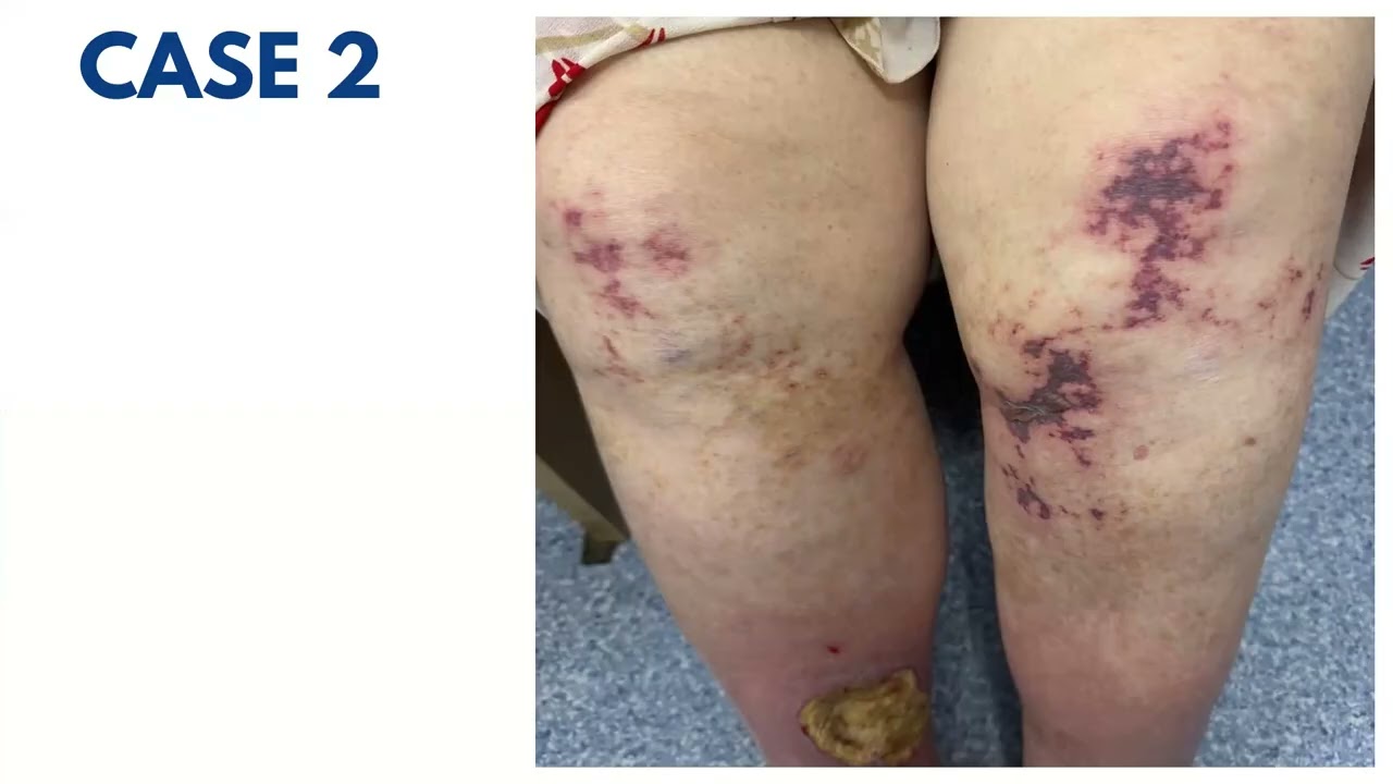
- Case 2: elderly patient with monoclonal gammopathy and connective tissue disease, annular/retiform purpura and ulceration, cryoglobulins positive.
- Cryoglobulinemic vasculitis types 1-3, association with hepatitis C, and the mixed small/medium vessel picture.
- ANCA-associated vasculitis: PR3 vs MPO, indirect immunofluorescence patterns, and the shift to antigen-specific testing.
- Granulomatosis with polyangiitis, microscopic polyangiitis and eosinophilic granulomatosis with polyangiitis with its three successive phases.
- Medium vessel vasculitis: polyarteritis nodosa and its cutaneous variant, hepatitis B association, and management.
- Brief note on large vessel vasculitis (giant cell arteritis, Takayasu arteritis) and concluding morphology-based summary.
Contribution & Novelties
The presentation does not report new data; its contribution is pedagogical. It consolidates a morphology-first approach to cutaneous vasculitis, using lesion pattern (palpable purpura, retiform purpura, subcutaneous nodules, livedo, digital necrosis) as the primary discriminator of vessel size, and it illustrates each category with cases from the unit’s own practice. The most useful synthesis is the explicit mapping between clinical morphology and the small, mixed, medium and large vessel categories, together with the reminder that cryoglobulinemic and ANCA-associated disease can straddle the small/medium boundary. It also usefully flags the shift in ANCA testing from indirect immunofluorescence to antigen-specific assays.
Pour aller plus loin :
- Chapel Hill nomenclature for vasculitis (Jennette et al., 2012 revised International Chapel Hill Consensus Conference) — the classification framework referenced in the talk; see the overview at https://en.wikipedia.org/wiki/Vasculitis
- ANCA-associated vasculitis — background on PR3/MPO serology and the ACR/EULAR 2022 classification criteria; see https://en.wikipedia.org/wiki/Anti-neutrophil_cytoplasmic_antibody
- Cryoglobulinemia — types I-III, hepatitis C association and mixed cryoglobulinemic vasculitis; see https://en.wikipedia.org/wiki/Cryoglobulinemia
- Leukocytoclastic vasculitis — histopathology (nuclear dust, fibrinoid necrosis) as described in the talk; see https://en.wikipedia.org/wiki/Leukocytoclastic_vasculitis
- Polyarteritis nodosa and hepatitis B association — see https://en.wikipedia.org/wiki/Polyarteritis_nodosa
184 words
Radar Profile
The profile is that of a solid clinical teaching review: information quantity and technical level are relatively high, reflecting a dense, terminology-rich walkthrough of an entire disease spectrum, while quality and overall reliability sit slightly lower because the content is didactic and unsourced. The gap between technical density and verifiability is the defining feature of this radar.